
QUALITY EYE CARE
Mostafa Optometry Professional Corporation
Dr. Abdel-Karim Mostafa
Optometrist
Comprehensive Eye Examination
A comprehensive eye examination is a thorough examination of the eye in which we perform different tests and procedures in order to assess the vision and evaluate the health of the eye. It includes, but is not limited to: history taking, visual acuity tests, stereo vision (3D vision), color vision, refraction, cover test, slit lamp examination, measurment of eye pressure, assessment of eye muscles, confrontation visual field, and checking the back of the eye. More details below.
What to expect from your visit

-
History Taking
A patient's history can identify the parts of the clinical examination that need special attention and indicate a need for particular additional investigations. History taking includes complaints, as well as personal, ocular, medical, family, social and drug history.
-
Visual Acuity Tests
Measuring the visual acuity is the first test performed in our eye exam. We measure the distance visual acuity by using a modern computer-based system to determine the smallest letters a patient can read on a standardized chart (Snellen chart) held 20 feet (6 meters) away. For near vision, we use a reading card to check the vision at a distance of 16 inches (40 cm).
Far Vision (distance glasses) : distance vision means visual acuity at 20 feet (6 meters) or more. Measurement of distance visual acuity is usually done using a computer-based program. This chart has letters or characters that can be identified by the person being examined. It can have the alphabet, numbers, objects or pictures.
Intermediate Vision (Computer glasses) : Generally refers to eyesight at approximately arm's length (About 20 to 26 inches / 50 to 65 cm), used by some patients who are 40 years old or older for tasks such as computer work and viewing a car's speedometer when driving.
Near Vision (Reading glasses) : near vision means eyesight used for reading and other close-up tasks, generally at a range of 12 to 16 inches (about 40 cm) from the eyes. Measurement of near visual acuity is usually done using a reading card. This chart has letters or characters that can be identified by the person being examined printed on it. It can have the alphabet, numbers, objects or pictures.


-
Stereo Vision (3-D Vision)
We use Titmus Fly stereotest to measure the perception of depth and 3-dimensional structure obtained on the basis of visual information deriving from two eyes. It reflects the integrity of binocular vision.

-
Refraction (glasses/contact lens prescription)

Group of tests for accurate measurement of prescription of the eyeglasses
and contact lenses. They include:
-
Retinoscopy test, by using the retinoscope.
-
Autorefraction using the computerized machine: autorefractometer.
-
Subjective refraction by using the phoropter.
Cycloplegic refraction is sometimes used to determine a person's refractive error by temporarily paralyzing the muscles of the eye by installing certain examination eye drops.
-
Color Vision Test
Using the Ishihara color vision test (PseudoIsochromatic Plate), the doctor is able to check whether the patient is color blind and the severity of the color blindness.


-
Slit Lamp Examination
The slit lamp is an instrument that provides a magnified, three-dimensional (3-D) view of the different parts of the eye. Using this instrument, we can examine your eyes, especially the eyelids, cornea, conjunctiva, sclera, iris, lens and the back of the eye. Often a yellow dye (fluorescein) is used to help examine the cornea and tear layer.
-
Tonometry (measuring eye Pressure)
Tonometry is an eye test that measures the pressure of the eyes, to determine whether or not a patient may be at risk for glaucoma, which causes damage to the eye's optic nerve.

-
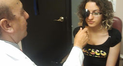
Cover Test
It is an objective determination of the presence and amount of ocular deviation (strabismus) or more subtle binocular vision problem (Phoria) that could cause the patient to suffer from eye strain.
-
Funduscopic Examination (examination of the back of the eye)
The interior surface of the eye (which includes the retina, optic disc, macula, fovea, and posterior pole) is an extremely important part of the eye's function. Mydriatic eye drops (such as tropicamide) may be used to dilate or enlarge the pupil in order to obtain a better view of the fundus of the eye.
